
CCAD is an allergic endoscopic phenotype—not a CT diagnosis
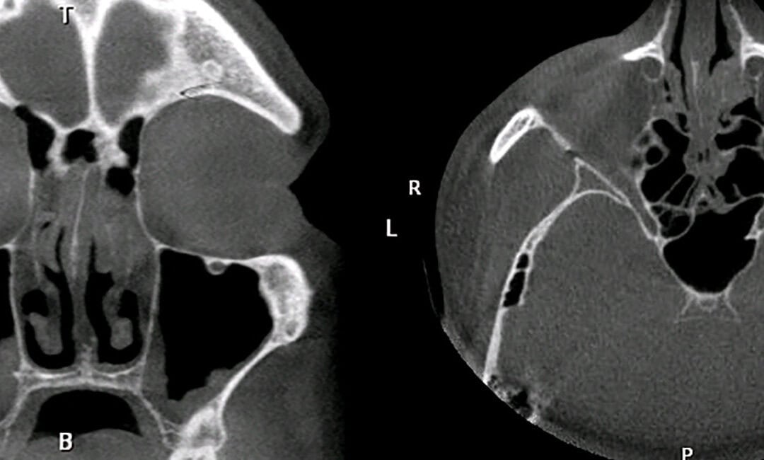
Central compartment atopic disease, or CCAD, is often recognised on a sinus CT scan. The central tissues of the nose become thickened while air remains around the outer margins of the sinuses, producing the characteristic “black halo” appearance.
It is an elegant radiologic sign. But it is not the disease.
CCAD is fundamentally a phenotype of the patient: usually a younger person with a clinical history consistent with inhalant allergy and visible allergic tissue remodelling arising from the central structures of the nose. The diagnosis rests on that history and the endoscopic origin of the abnormal tissue. CT supports the diagnosis, but should not define it.
Correctly identifying CCAD changes the long-term treatment strategy, including the role of allergen immunotherapy, surgery and, in severe disease, biologic therapy.
What is central compartment atopic disease?
CCAD is a form of primary diffuse type 2 chronic rhinosinusitis associated with inhalant allergy.[1–3] The allergic response produces oedema and gradual tissue remodelling in the parts of the nose that receive concentrated exposure to inhaled particles:
- the middle turbinate
- the superior turbinate
- the posterosuperior nasal septum
- sometimes the adjacent uncinate region
The earliest change may be quite subtle: smooth oedema arising from the middle turbinate. With continued allergic inflammation, that tissue may become multifocal or develop into discrete central polyps.
A 2026 international Delphi consensus proposed an endoscopic grading system that captures this spectrum.[10] Grade 1 is middle turbinate or posterosuperior septal oedema without polyps. Grade 2 describes small polyps arising from the middle or superior turbinate. Grade 3 includes polyps medial to the middle turbinate, usually arising from the posterosuperior septum. Grade 4 means that one of these CCAD patterns coexists with diffuse polyps arising from the middle meatus. It is a mixed phenotype, not simply a more advanced stage of pure CCAD.
This is why a nasal polyp is not a diagnosis . The important question is not simply whether polypoid tissue is present, but where it arose and why it developed.
The defining feature is central tissue remodelling on endoscopy
The most important diagnostic step is nasal endoscopy. In CCAD, the abnormal tissue arises from the turbinates or upper septum. Middle turbinate oedema typically has a smooth, semitranslucent, almost jelly-like appearance.[3,4]
This differs from typical diffuse eosinophilic chronic rhinosinusitis with nasal polyps, where “true” polyps emerge from within the middle meatus or sinus cavity. The two conditions can share type 2 inflammation and tissue eosinophilia, but the anatomical origin of the remodelling is different.[4,5]

Inferior turbinate hypertrophy, cobblestoning, a pseudoturbinate along the nasal floor and clear allergic secretions provide further endoscopic evidence of an allergic nose. These findings are more informative than labelling every smooth polypoid structure as generic “nasal polyposis.”
The clinical history is part of the phenotype
CCAD is not only something seen through an endoscope. The patient usually has a recognisable clinical history.
The allergic symptoms often begin well before the sinus disease. Patients commonly describe sneezing, itching, watery rhinorrhoea, allergic eye symptoms and regular antihistamine use. They are often younger than patients with classic diffuse eosinophilic polyposis.[2,4,6]
Our recent study examined 91 patients with endoscopically unambiguous type 2 disease: 23 with CCAD and 68 with eosinophilic CRS. Clinical-history features alone, analysed without the endoscopic diagnosis, reproduced the expert-assigned phenotype in 93% of patients. A history-only model discriminated the two groups with a cross-validated area under the curve of 0.90.[7]
The characteristic pattern was not simply “has allergy”:
- CCAD patients were younger and much more likely to report clear allergy symptoms and regular antihistamine use.
- Asthma, when present, was more likely to have begun in childhood. By adulthood, upper-airway disease could dominate while asthma was relatively quiet.
- Barotrauma—sinus pain with flying, diving or marked atmospheric-pressure change—was considerably more common.
- Early smell loss, synchronous upper- and lower-airway onset and early postoperative polyp recurrence were less characteristic than in diffuse eosinophilic disease.
These findings need external validation, particularly in consecutive patients and mixed disease. They should complement rather than replace endoscopy. However, they reinforce the central point: CCAD represents a distinct allergic patient phenotype, not merely a pattern drawn on a CT scan.
What defines CCAD—and what does not?
| Finding | Diagnostic role |
|---|---|
| Allergic clinical history, often beginning years earlier | Strongly supports CCAD and identifies the patient phenotype |
| Polypoid thickening or discrete polyps arising from the middle or superior turbinate or posterosuperior septum | Defining endoscopic feature |
| Isolated middle turbinate or posterosuperior septal oedema without polyps | Strong allergic marker and Grade 1 CCAD in the 2026 consensus system |
| Inferior turbinate hypertrophy, cobblestoning or pseudoturbinate formation | Supportive signs of an allergic nose |
| Central thickening with peripheral sinus air—the “black halo” sign | Characteristic and confirmatory, but not diagnostic by itself |
| Positive skin-prick or serum-specific IgE testing | Helpful when it matches the history, but not essential in every patient |
| Negative systemic allergy testing | Does not exclude CCAD or local nasal allergy |
| Polyps arising from the middle meatus or sinus cavity | More consistent with eosinophilic CRS or mixed disease than pure CCAD |
| Diffuse sinus opacification in advanced disease | Does not exclude CCAD because secondary mucus trapping can obscure the original central pattern |
Why the black halo sign is not the definition
Central turbinate enlargement may narrow or obstruct the sinus drainage pathways. The central tissues therefore appear opaque on CT while the more peripheral sinus mucosa remains relatively normal. This creates the “black halo” of air surrounding the central disease.[2,6]

Coronal and axial CT images showing central nasal thickening with peripheral sinus aeration—the classic black halo pattern associated with CCAD.
The sign is highly characteristic when present, but CT can fail at both ends of the disease spectrum. Early remodelling may be too subtle to classify. In advanced CCAD, central tissues obstruct the sinus openings, mucus becomes trapped and peripheral opacification may erase the original halo. Mixed disease creates a further problem because genuine central remodelling and sinus-derived polyps can coexist.
Some published studies have nevertheless assigned CCAD solely from radiologic appearances. That is a methodological error. A “central radiologic phenotype” may be useful for describing a scan, but it is not interchangeable with an endoscopically confirmed clinical diagnosis.
The problem is clearest when a study requires the black halo and excludes advanced pansinus disease. It has selected classifiable CT scans while removing cases in which progressive CCAD has lost its classic appearance.[11] Such findings may be valid for that CT pattern, but should not redefine CCAD.
Recent literature synthesis and international expert consensus are clear: endoscopic confirmation of central compartment remodelling is critical, while radiology alone is insufficient.[1,10]
The phenotype may also vary geographically with persistent inhalant allergy and local allergen exposure. Inconsistent or radiology-only definitions can exaggerate those differences.
A negative allergy test does not end the assessment
Skin-prick and serum-specific IgE testing remain useful, particularly for a perennial allergen such as house dust mite. But sensitisation is not identical to clinically active allergy, and allergen-specific IgE has been demonstrated within central tissue when the corresponding skin and serum tests were negative.[8] History, endoscopy and testing must be interpreted together.
This is the same principle discussed in when a negative allergy test does not rule out allergy .
Why identifying CCAD changes treatment
Phenotyping matters only if it leads to more rational care. In CCAD, treatment should address both the structural consequence and the allergic driver.
Topical corticosteroids remain useful for suppressing inflammation, and surgery may be necessary when established turbinate remodelling causes nasal or sinus obstruction. Surgery can remove the remodelled tissue, reopen the sinuses and improve delivery of topical treatment. It does not, however, change the underlying inhalant-allergy response.
For a patient who has progressed to surgery despite appropriate pharmacotherapy, returning to the same corticosteroid-only strategy that failed to prevent progression is incomplete. The structural disease has been treated, but the disease mechanism has not.
That is where allergen immunotherapy becomes important. In our retrospective cohort of 86 surgically treated, house dust mite-sensitised CCAD patients, 32 received allergen immunotherapy. At 12 months, diffuse or polypoid middle turbinate oedema was present in 15.6% of the immunotherapy group compared with 52.9% of those not receiving immunotherapy. Frequent corticosteroid-irrigation use was also lower, although symptom scores were similar.[9]
This retrospective study is not definitive proof, but it provides CCAD-specific evidence that modifying the allergic response may reduce recurrent tissue activity and allow de-escalation of topical treatment.
This is why immunotherapy should not be the last step in allergic rhinitis , particularly in a young patient already showing allergic tissue remodelling.
What about biologic therapy?
Some patients with severe, uncontrolled type 2 disease may ultimately require biologic treatment. The phenotype remains relevant here.
Classic CCAD is driven by inhalant allergy and local allergic remodelling rather than being defined simply by a high circulating eosinophil count. CCAD tissue can express IL-5 and IL-13, but published comparisons also suggest a lower overall eosinophilic inflammatory burden than in classic eCRS.[5,11]
In our clinical experience, anti-IL-5 therapy is often less convincing in this allergy-dominant phenotype than treatment blocking IL-4 and IL-13 signalling. However, there are no head-to-head CCAD trials and the existing evidence does not prove that one class is superior.
Biologic selection should consider the whole patient: endoscopic phenotype, clinically relevant allergy, asthma pattern, blood and tissue markers, other type 2 disease and previous treatment response. CCAD should inform selection, not become an unsupported shortcut.
Conclusion
CCAD is not defined by a black halo on CT. It is defined by central turbinate and septal tissue remodelling in a patient with a clinical phenotype of inhalant allergy.
The black halo is a consequence of that process and may be striking, subtle or lost as disease advances. The reliable hierarchy is history, endoscopic confirmation, allergy assessment and then CT interpreted in that context.
Recognising this phenotype supports early treatment of the allergic driver, avoids relying on corticosteroids alone after structural disease has progressed, strengthens the role of immunotherapy and provides a more rational framework for surgery and biologics.
The scan is helpful. The patient and the endoscopy define the disease.
References
- Barrow EM, Wise SK, Botros A, Harvey RJ, Edwards TS, DelGaudio JM. Central compartment atopic disease: analysis of current literature and proposal of diagnostic criteria. Int Forum Allergy Rhinol. 2025;15(10):1113–1135. doi:10.1002/alr.70012
- DelGaudio JM, Loftus PA, Hamizan AW, Harvey RJ, Wise SK. Central compartment atopic disease. Am J Rhinol Allergy. 2017;31(4):228–234. doi:10.2500/ajra.2017.31.4443
- Hamizan AW, Christensen JM, Ebenzer J, et al. Middle turbinate edema as a diagnostic marker of inhalant allergy. Int Forum Allergy Rhinol. 2017;7(1):37–42. doi:10.1002/alr.21835
- Brunner JP, Jawad BA, McCoul ED. Polypoid change of the middle turbinate and paranasal sinus polyposis are distinct entities. Otolaryngol Head Neck Surg. 2017;157(3):519–523. doi:10.1177/0194599817711887
- Sit A, Alvarado R, Earls P, et al. CCAD or eCRS: defining eosinophilic subpopulations in chronic rhinosinusitis. Am J Rhinol Allergy. 2023;37(4):402–409. doi:10.1177/19458924231155012
- Hamizan AW, Loftus PA, Alvarado R, et al. Allergic phenotype of chronic rhinosinusitis based on radiologic pattern of disease. Laryngoscope. 2018;128(9):2015–2021. doi:10.1002/lary.27180
- Seresirikachorn B, Alvarado R, Perkovic A, et al. Clinical history discriminates endoscopically defined central compartment atopic disease from eosinophilic chronic rhinosinusitis. Manuscript submitted for publication. 2026.
- Edwards TS, DelGaudio JM, Levy JM, Wise SK. A prospective analysis of systemic and local aeroallergen sensitivity in central compartment atopic disease. Otolaryngol Head Neck Surg. 2022;167(5):885–890. doi:10.1177/01945998221082554
- Meerwein CM, Sacks PL, Ho J, et al. The effect of allergen immunotherapy in patients with central compartment atopic disease post-surgery. Int Forum Allergy Rhinol. 2025;15(2):128–134. doi:10.1002/alr.23459
- Moreno-Luna R, Palma-Martínez C, Sánchez-Gómez S, et al. Rationale of new grading system: central compartment atopic disease. Int Forum Allergy Rhinol. 2026;16(7):670–680. doi:10.1002/alr.70114
- Lin YT, Lin CF, Liao CK, Chiang BL, Yeh TH. Clinical characteristics and cytokine profiles of central-compartment-type chronic rhinosinusitis. Int Forum Allergy Rhinol. 2021;11(7):1064–1073. doi:10.1002/alr.22759
Frequently asked questions
What is central compartment atopic disease?
Central compartment atopic disease, or CCAD, is an allergy-associated form of chronic rhinosinusitis in which oedema, polypoid change or polyps arise from central nasal structures such as the middle or superior turbinates and the posterosuperior septum. It is defined by the allergic clinical phenotype together with this central tissue remodelling on endoscopy.[1–3,10]
Is the black halo sign diagnostic of CCAD?
No. Central thickening with peripheral sinus aeration creates the characteristic black halo appearance on CT and strongly supports the diagnosis. However, it is a consequence of the disease rather than its defining feature, and radiology alone is insufficient to diagnose CCAD.[1,2,10]
What does CCAD look like on nasal endoscopy?
The earliest finding may be smooth oedema of the middle turbinate or posterosuperior septum. More established disease can produce polypoid remodelling or discrete polyps arising from the middle or superior turbinate or upper septum, rather than true sinus polyps emerging from the middle meatus.[3,4,10]
Can advanced CCAD lose the black halo appearance?
Yes. Progressive central remodelling can obstruct sinus drainage, trap mucus and produce more diffuse sinus opacification. The original black halo pattern may therefore become obscured, particularly in advanced or mixed disease.[1,10]
Does Grade 4 CCAD simply mean more severe pure CCAD?
No. In the 2026 consensus grading system, Grade 4 describes a mixed phenotype in which central CCAD changes coexist with diffuse polyps arising from the middle meatus. It is not merely the most advanced stage of pure CCAD.[10]
Can CCAD occur despite negative conventional allergy tests?
Yes. Negative skin-prick or serum-specific IgE tests do not automatically exclude clinically relevant allergy or a local allergic response within the nose. The history, endoscopic findings and allergy testing should be interpreted together.[8]
Why does identifying CCAD change treatment?
Surgery can remove established remodelling and reopen the sinuses, but it does not change the underlying inhalant-allergy response. Recognising CCAD supports treatment directed at the allergic driver, including allergen avoidance and immunotherapy when appropriate, alongside topical treatment, surgery and carefully selected biologic therapy.[9]