
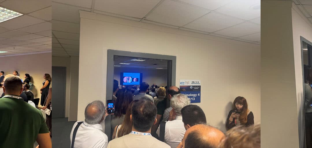
At the recent IFOS meeting, the interest in septal perforation was difficult to miss.
Sessions devoted to septal perforation reconstruction were standing room only. Colleagues were crowded into the room, filling the doorway and spilling into the corridor.
That interest reflects how much this field has changed.
For many years, the principal question after septal perforation surgery was simple:
Did the hole close?
That remains important. But contemporary septal perforation surgery increasingly asks a better question:
Did we reconstruct a healthy, comfortable and stable nasal septum?
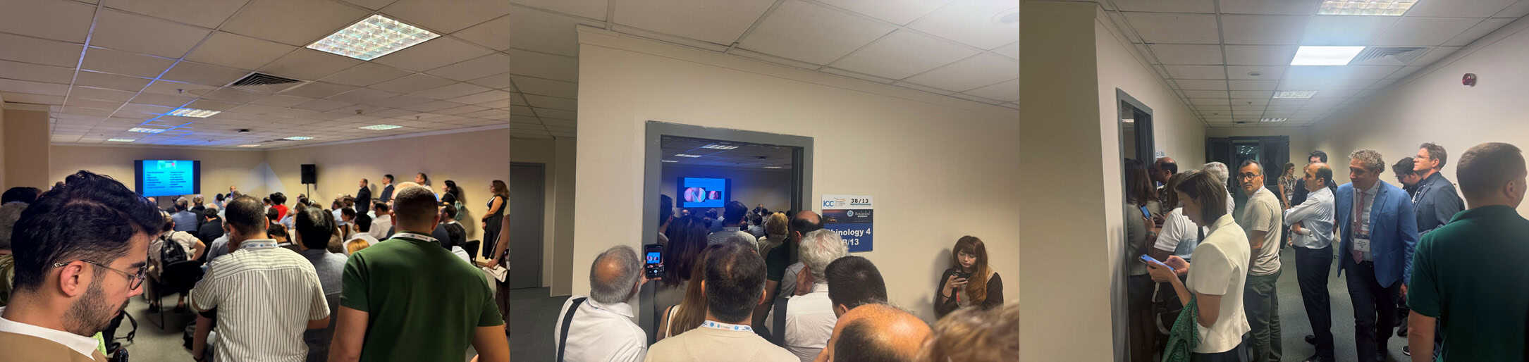
Standing room only at a septal perforation session at IFOS 2026. The level of interest reflects the rapid evolution in septal perforation reconstruction and postoperative healing.
Closing the hole is only one outcome
The recently published International Consensus Statement on Nasal Septal Perforation, which I was involved in as a senior author, provides a useful marker of how thinking has evolved.[1]
One of its important conclusions is that surgical closure and clinical success are not necessarily the same thing.
A perforation can be completely closed but the patient may continue to experience crusting, bleeding, irritation or obstruction. The consensus therefore considers both anatomical and clinical outcomes when defining therapeutic success.[1]
Complete success means a closed perforation without symptoms. A closed but symptomatic nose represents surgical success without clinical success.
That distinction sounds simple, but it represents an important change in how septal perforation surgery should be judged.
The perforation is not simply a hole. Crusting, desiccation, ulceration and repeated trauma can perpetuate mucosal injury around its margins.[1] The quality of the lining covering the reconstructed septum therefore matters.
For patients wanting to understand the broader principles of repairing particularly large defects, see Large Septal Perforations: Why “Too Big to Fix” Is No Longer the Right Answer.
The goal is healthy mucosa
An increasingly important reconstructive principle is to provide healthy mucosal coverage wherever possible—ideally on both sides of the repair.
The international consensus specifically supports this concept. Providing mucosal coverage on both sides was considered advisable, and contralateral free mucosal grafts or interposition grafts can be used with a pedicled flap in selected cases.[1]
There are several ways of achieving this.
One is to preserve useful mucosa already present around the perforation.
My colleague Professor Isam Alobid and his Barcelona group have developed elegant techniques such as the Boot-on-Donut and Racket-on-Donut, where suitable perforation edges are everted so that this existing mucosa helps resurface the opposite side of the reconstruction rather than simply being discarded.[2,3]
This illustrates an important change in philosophy: the perforation edge can sometimes be useful reconstructive tissue.
Vascularised flaps have expanded what can be repaired
Vascularised mucosal flaps remain the foundation of many contemporary repairs.
The anterior ethmoidal artery flap has become an important technique for endoscopic septal reconstruction, but previous surgery may leave traditional septal mucosa scarred, damaged or unavailable.
Alternative sources of vascularised mucosa have therefore become increasingly important.
One example is the Boxer’s flap, developed by Yury Rusetsky. This is an anteriorly pedicled nasal floor flap based on the superior labial artery and provides another reconstructive option when more traditional septal flaps are not feasible.[4]
The recently published series reported anatomical closure in 90.5% of 21 patients, together with substantial symptomatic improvement.[4] The publication also deliberately assessed complete success using both closure and symptom resolution.
Free mucosal grafting: resurfacing the other side
The next question is what happens on the opposite side of the reconstruction.
A vascularised flap can provide excellent coverage on one side of the septum, but the contralateral surface may otherwise be left to heal slowly by secondary epithelialisation.
Our research group has therefore been using a relatively simple adjunct: a free mucosal graft placed on the contralateral surface.
The graft is usually harvested from the inferior turbinate, thinned and spread over the opposite surface of the repair. It does not replace the vascularised flap. The vascularised flap remains the foundation of the reconstruction.
The purpose of the free graft is different: to establish healthy mucosa on the opposite surface as early as possible.
Nick Campion and our research group have now evaluated this approach in 90 patients in Free Mucosal Grafting is a Simple Adjunct for Rapid Re-Epithelization in Septal Perforation Repair, now in press in Laryngoscope.[5]
Anatomical closure was achieved in 90% of patients.
Among the repairs achieving complete anatomical closure, 92.6% demonstrated complete integration of the free mucosal graft.[5]
Nasal symptoms and endoscopic appearances also improved significantly.[5]
This is important because a thin free mucosal graft placed over the reconstruction might intuitively seem unlikely to survive reliably. The results suggest otherwise.
This retrospective case series had no ungrafted control group, so it supports the feasibility and reliable integration of free mucosal grafting rather than proving superiority over healing without a graft.[5]
Rather than leaving a relatively large surface to heal slowly from its margins, free grafting provides another means of trying to restore normal mucosa early.
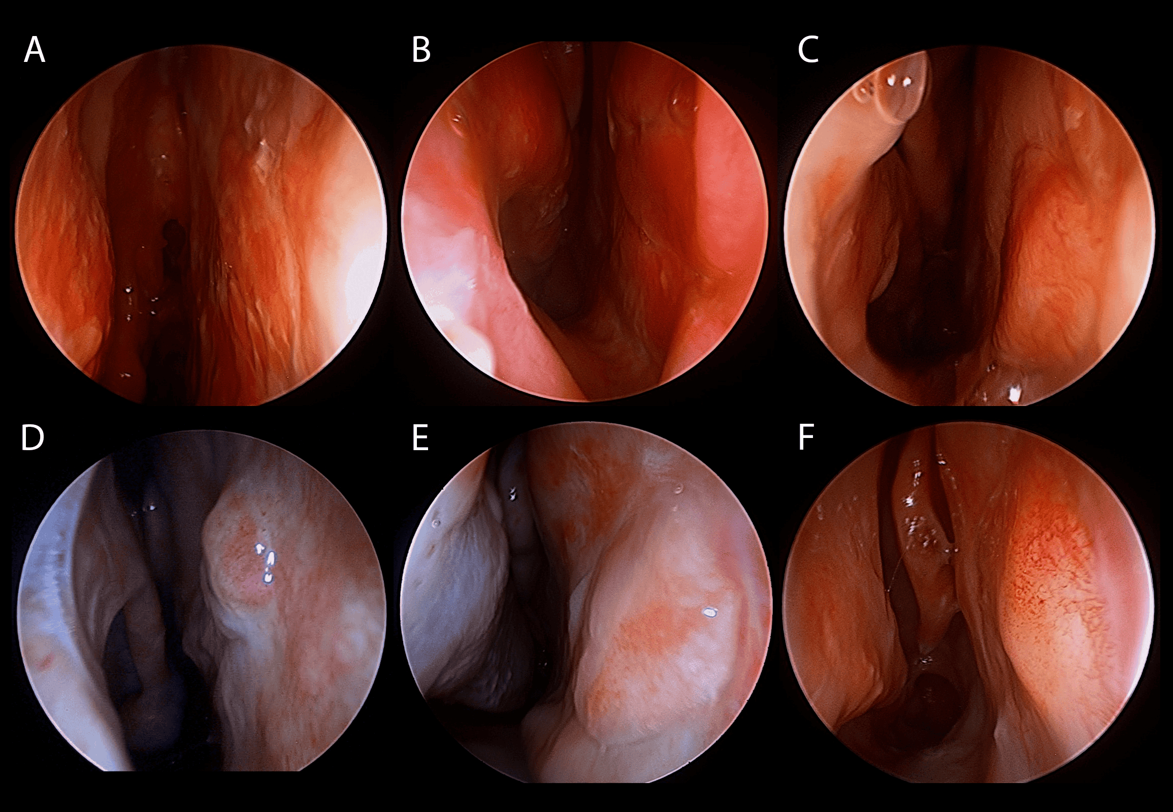
Healthy mucosal healing after free mucosal grafting. The upper examples demonstrate early graft integration at approximately six weeks following removal of the silicone sheets, while the lower examples demonstrate stable mucosal healing at later postoperative time points.
Silicone sheets may matter more than we once thought
The postoperative period should also be considered part of the reconstruction rather than simply recovery from the operation.
Silicone—or Silastic—sheets protect the reconstructed surfaces from drying, mechanical trauma and shear while healing occurs.
The international consensus recommends silicone splints on both sides of the septum for at least two weeks after surgery.[1] In the consensus process there was considerable variation in duration, although most experts reported leaving them in place for approximately three to four weeks.[1]
In our free-graft technique, we generally leave them for four to six weeks.[5]
We use very thin 0.5 mm silicone sheets, individually shaped from 5 × 5 cm sheets to fit the reconstructed septum.
Their role is not simply to hold the septum in position.
They provide a protected, moist environment while the mucosal surfaces establish themselves. Experimental and clinical work suggests that protection from desiccation and mechanical disruption assists septal mucosal regeneration.[5]
Our current study cannot prove that four to six weeks is superior to earlier removal, because all patients were treated with the same protocol. But it reinforces an important principle:
The operation creates the reconstruction; the healing environment helps determine its final quality.
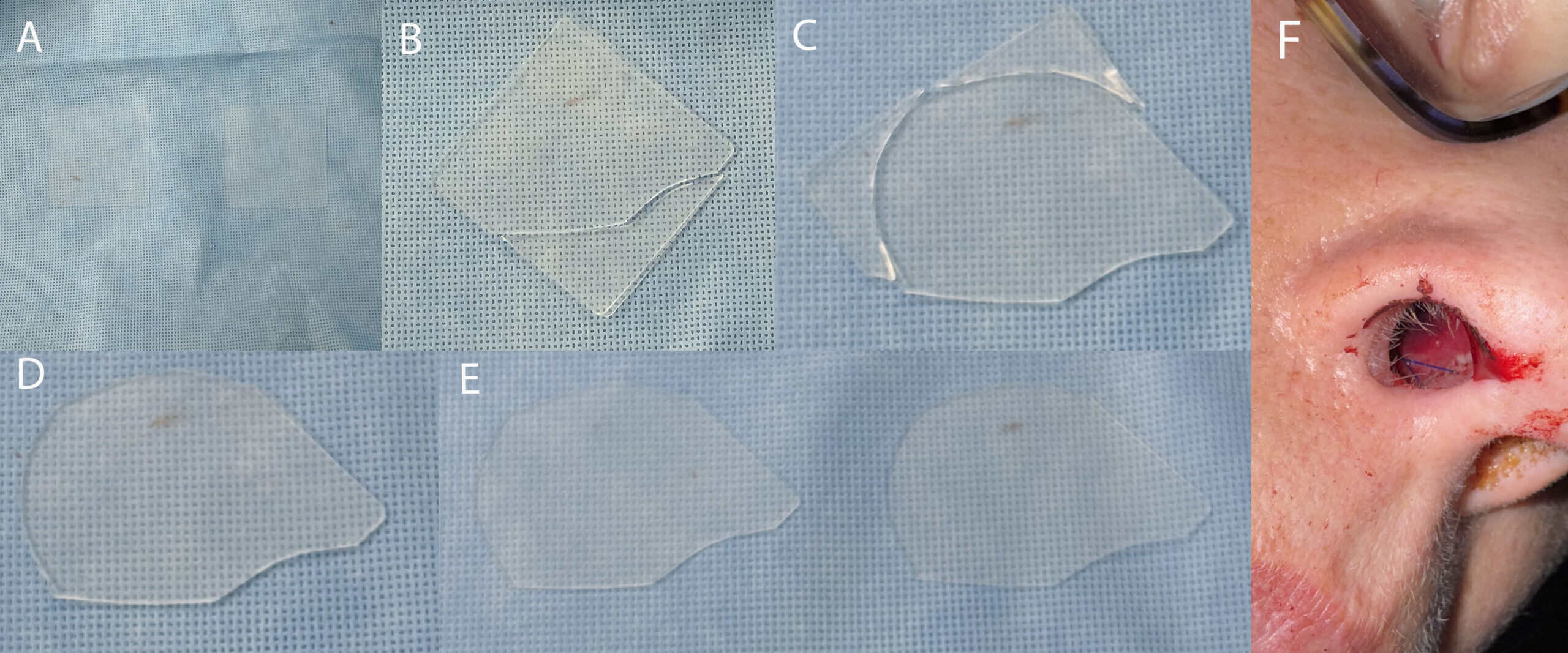
Custom-shaped silicone sheets used after septal perforation reconstruction. Thin 0.5 mm sheets are cut from 5 × 5 cm silicone sheets and shaped individually to protect the reconstructed septal surfaces during healing.
The field itself has matured
The expansion of specialist education around septal perforation is another indication of how rapidly the field is developing.
Professor Isam Alobid directs the International Septal Perforation Repair “State-of-the-art” Course in Barcelona. Its program includes anterior ethmoidal, greater palatine, nasal floor and other vascularised flaps, together with Boot-on-Donut, Racket-on-Donut and other reconstructive techniques.
Isam is also editor of the Springer text Septal Perforation Repair: State-of-the-Art, a dedicated monograph on septal perforation repair.
The crowded sessions at IFOS make more sense in that context.
Septal perforation used to be regarded as a frustrating condition with unpredictable surgery and relatively limited reconstructive options.
Today we have vascularised endoscopic flaps, alternative donor sites, everted-edge techniques, interposition materials, free mucosal grafting and a much better appreciation of postoperative mucosal healing.
The question is no longer simply:
Can we close the perforation?
It is:
Can we reconstruct a healthy septum that stays closed, heals with good mucosa and actually makes the patient feel better?
That is a much higher standard. And it is increasingly the standard by which septal perforation repair should be judged.
References
- Alobid I, Garaycochea O, Rojas-Lechuga MJ, et al. International consensus statement on nasal septal perforation. Rhinology. 2026. doi:10.4193/Rhin26.027.
- Alobid I, Bernal-Sprekelsen M. “Boot-on-Donut” procedure for anterior and large septal perforation. Eur Arch Otorhinolaryngol. 2024;281:2761–2765.
- Alobid I, Santamaría-Gadea A, Mariño-Sánchez F. Endoscopic “Racket-on-Donut” technique for large anterior nasoseptal perforations. Laryngoscope. 2024;134:143–147.
- Campion NJ, Mokoyan Z, Stepanova V, Alobid I, Harvey RJ, Rusetsky Y. Superior labial artery flap for septal perforation repair: the Boxer’s flap. Laryngoscope. 2026. doi:10.1002/lary.70690.
- Campion NJ, Couto AM, Hua E, Qiao J, Gendeh H, Rosenbaum Fuentes A, et al. Free Mucosal Grafting is a Simple Adjunct for Rapid Re-Epithelization in Septal Perforation Repair. Laryngoscope. In press.
Frequently asked questions
Is closing a septal perforation enough to make the operation successful?
Not always. Modern assessment considers both whether the perforation closes and whether symptoms such as crusting, bleeding, irritation and obstruction improve. A technically closed perforation that remains symptomatic is not the same as complete therapeutic success.
Why is healthy mucosa important after septal perforation repair?
Healthy mucosa provides the normal lining of the nasal septum. Areas left raw or poorly epithelialised are more susceptible to drying, crusting and irritation. Contemporary techniques increasingly aim to restore healthy mucosal coverage on both sides of the repair.
What is a free mucosal graft?
A free mucosal graft is a thin piece of healthy nasal lining harvested from another area of the nose and transferred to the reconstruction. In this technique it is placed opposite a vascularised flap to assist rapid resurfacing of the contralateral side.
How long are silicone sheets left in after septal perforation repair?
There is no single universally agreed duration. International consensus supports bilateral silicone splints for at least two weeks, while many experts use approximately three to four weeks. In the free mucosal graft technique described here, silicone sheets are generally left for four to six weeks.
Can large septal perforations still be repaired?
Size makes septal perforation reconstruction more difficult, but size alone does not determine whether repair is possible. The availability and quality of healthy vascularised mucosa, previous surgery, mucosal condition and perforation location are also important.